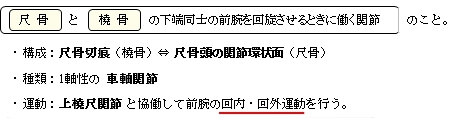
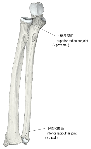
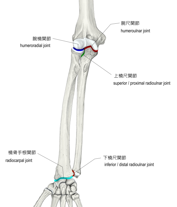
「The distal radioulnar articulation (inferior radioulnar joint) is a joint between the two bones in the forearm; the radius and ulna. It is one of two joints between the radius and ulna, the other being the proximal radioulnar articulation. The distal radioulnar articulation is the one of the two closest to the wrist and hand.
The distal radioulnar articulation pivot-joint formed between the head of ulna and the ulnar notch on the lower extremity of radius.
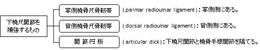
Ligaments:The articular surfaces are connected together by the following ligaments:
Function:The function of the radioulnar joint is to lift and maneuver weight load from the distal radioulnar joint to be distributed across the forearm’s radius and ulna as a load-bearing joint.[1]Supination of the radioulnar joint can move from 0 degrees neutral to approximately 80-90 degrees where Pronation of the Radioulnar Joint can move from 0 degrees neutral to approximately 70-90 degrees.[2] Supination (palms facing up) vs. pronation (palms facing down). Muscles that contribute to function are all supinator (Biceps Brachii, Brachioradialis, and Supinator) and pronator muscles (Brachioradialis, Pronator Quadratus, and Pronator Teres).」