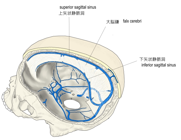
「The falx cerebri is also known as the cerebral falx, named from its sickle(かま)-like form. It is a large, crescent-shaped fold of meningeal layer of dura mater that descends vertically in the longitudinal fissure between the cerebral hemispheres. The falx cerebri attaches anteriorly at the crista galli in proximity to the cribriform plate and to the frontal and ethmoid sinuses. Posteriorly, it is connected with the upper surface of the tentorium cerebelli. Its superior margin is attached at midline to internal surface of skull, as far back as the internal occipital protuberance. The superior sagittal sinus is contained in the superior margin of the falx cerebri and overlies the longitudinal fissure of the brain. The inferior sagittal sinus is contained in the inferior margin of the falx cerebri and arches over the corpus callosum, deep in the longitudinal fissure. 」