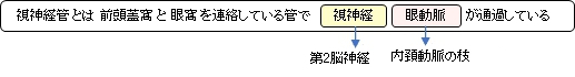
・「(小翼は)前根と後根の2根を有し、この両根の間に視神経管がある。」(船戸和也のHP)
・=視神経孔
管状といっても短いため(長さは不明)「孔(foramen)」と捉える資料も多く、その場合は「視神経管=視神経孔」としている。
ただ、「Wikipedia」では管と孔を分けている解説が見られる。
「The optic foramen is the opening to the optic canal. 」
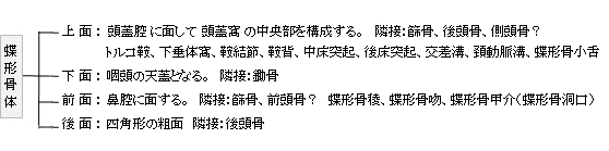
・「(視神経交叉溝は )鞍結節の前方を横走する溝、視神経交叉を入れ、その両端は視神経管に続いている。」(日本人体解剖)
|
|
|
|
視神経管 |
右眼窩(横断面) |
視交叉 |
頭蓋窩 |
|
|
|
|
蝶形骨(上面) |
眼窩裂、視神経孔 |
|
|
以下は「Wikipedia」の解説文となる。
The optic foramen is the opening to the optic canal. The canal is located in the sphenoid bone; it is bounded medially by the body of the sphenoid and laterally by the lesser wing of the sphenoid.
The superior surface of the sphenoid bone is bounded behind by a ridge, which forms the anterior border of a narrow, transverse groove, the chiasmatic groove (optic groove), above and behind which lies the optic chiasma; the groove ends on either side in the optic foramen, which transmits the optic nerve and ophthalmic artery (with accompanying sympathetic nerve fibres) into the orbital cavity. Compared to the optic nerve, the ophthalmic artery is located inferolaterally within the canal.
The left and right optic canals are 25mm apart posteriorly and 30mm apart anteriorly. The canals themselves are funnel-shaped (narrowest anteriorly).
【 語 句 】
・optic foramen:視神経孔 ・sphenoid bone:蝶形骨 ・lesser wing:小翼 ・ridge:隆起(線) ・groove:溝 ・chiasmatic groove:視神経交差溝 ・optic chiasma:視交叉 ・optic nerve:視神経 ・ophthalmic artery:眼動脈 ・sympathetic nerve:交感神経 ・orbital cavity:眼窩
■ 写真やイラストを掲載しているサイト ■
・ イラストや写真を掲載しているサイト-Ⅰ
・ イラストや写真を掲載しているサイト-Ⅱ
・ イラストや写真を掲載しているサイト-Ⅲ
・ イラストや写真を掲載しているサイト-Ⅳ
・ イラストや写真を掲載しているサイト-Ⅴ