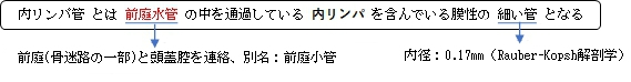
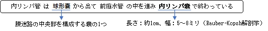
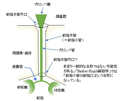
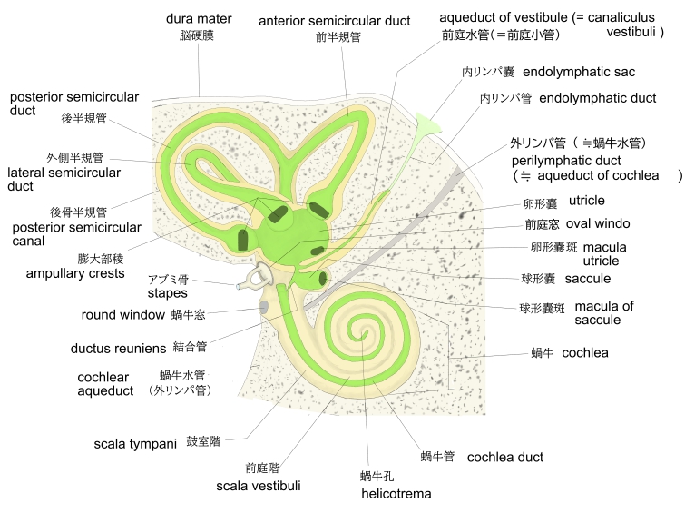
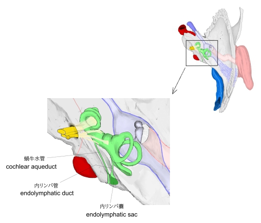
内リンパ管( ないりんぱかん、英:endolymphatic duct ) 連嚢管~内リンパ嚢 (模型図) 骨迷路と膜迷路 右内耳 (上方から見て) 以下は「Rauber-Kopsh解剖学」の解説文となる。 「卵形嚢は球形嚢の壁と1ヵ所で寄り合っている.といっても両者の壁が融合して1枚の壁に,つまり1枚の隔壁になっているというわけではなく,両者の壁はたがいに分離している.内径0.17mmの細長い1本の管が両嚢と開放性に結合しており,これを内リンパ管Ductus endolymphaceusという.内リンパ管から球形嚢につづく脚の方が太くて,もう1つの卵形嚢に開く脚の方が細い.後者を連嚢管Ductus utriculosaccularisという.内リンパ管はその末端部でふくらんで,内リンパ嚢Saccus endolymphaceusというかなりの大きさの平たいふくろになっている.これは長さ約1cm,幅5~8mmのふくろで,前庭小管の内口より外側にあり,側頭骨の錐体乳突部の後面に接して硬膜の2葉のあいだにはさまれている.その方向は下外側方へと伸びている. 」 また、「Wikipedia」では以下のように解説している。 From the posterior wall of the saccule a canal, the endolymphatic duct, is given off; this duct is joined by the ductus utriculosaccularis, and then passes along the aquaeductus vestibuli and ends in a blind pouch (endolymphatic sac) on the posterior surface of the petrous portion of the temporal bone, where it is in contact with the dura mater. Disorders of the endolymphatic duct include Meniere's Disease and Enlarged Vestibular Aqueduct. 【 語 句 】 ・saccule:球形嚢 ・ductus utriculosaccularis:連嚢管 ・aquaeductus vestibuli:前庭水管 ・blind pouch:盲管 ・endolymphatic sac:内リンパ嚢 ・petrous portion of the temporal bone:側頭骨の錐体部 ・dura mater:硬膜 ・Meniere's Disease:メニエール病 ・Enlarged Vestibular Aqueduct:前庭水管拡大 ■ 写真やイラストを掲載しているサイト ■ ・ イラストや写真を掲載しているサイト-Ⅰ ・ イラストや写真を掲載しているサイト-Ⅱ ・ イラストや写真を掲載しているサイト-Ⅲ ・ イラストや写真を掲載しているサイト-Ⅳ ・ イラストや写真を掲載しているサイト-Ⅴ