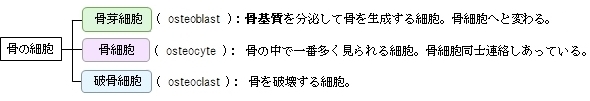
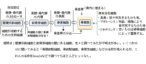
「An osteocyte, a star-shaped type of bone cell, is the most commonly found cell in mature bone tissue, and can live as long as the organism itself.[1] The adult human body has about 42 billion osteocytes.[2] Osteocytes have an average half life of 25 years, they do not divide, and they are derived from osteoprogenitors, some of which differentiate into active osteoblasts.[1] Osteoblasts/osteocytes develop in mesenchyme.
In mature bone, osteocytes and their processes reside inside spaces called lacunae (Latin for a pit) and canaliculi, respectively.[1] When osteoblasts become trapped in the matrix that they secrete, they become osteocytes. Osteocytes are networked to each other via long cytoplasmic extensions that occupy tiny canals called canaliculi, which are used for exchange of nutrients and waste through gap junctions.
Although osteocytes have reduced synthetic activity and (like osteoblasts) are not capable of mitotic division, they are actively involved in the routine turnover of bony matrix, through various mechanosensory mechanisms. They destroy bone through a rapid, transient (relative to osteoclasts) mechanism called osteocytic osteolysis. Hydroxyapatite, calcium carbonate and calcium phosphate is deposited around the cell.」