「日本人体解剖学 (上巻) 」では以下のように解説している。 」では以下のように解説している。
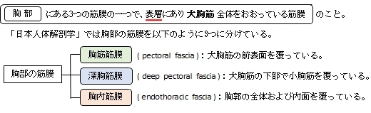
「大胸筋の前表面をおおい、特に内側上方が発達し三角筋(大)胸筋溝で深胸筋膜と合する。上方は頚部の筋膜の浅葉に、下方は浅腹筋膜に、外側方は三角筋筋膜および腋窩筋膜に移行し、内側方は胸骨につく。 」
それに対して、「Rauber-Kopsch解剖学」で「胸壁の筋膜」を以下のように3つに分けている。
「胸壁には3つの筋膜がある,すなわち浅胸筋膜,鎖骨胸筋膜および胸内筋膜である.
つまり、ここでいう「浅胸筋膜」が「日本人体解剖学 (上巻) 」の「胸筋筋膜」だと思われる。
また、以下は「Wikipedia」の解説文となる。
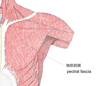
「The pectoral fascia is a thin lamina, covering the surface of the pectoralis major, and sending numerous prolongations between its fasciculi: it is attached, in the middle line, to the front of the sternum; above, to the clavicle; laterally and below it is continuous with the fascia of the shoulder, axilla, and thorax.
It is very thin over the upper part of the pectoralis major, but thicker in the interval between it and the latissimus dorsi, where it closes in the axillary space and forms the axillary fascia; it divides at the lateral margin of the latissimus dorsi into two layers, one of which passes in front of, and the other behind it; these proceed as far as the spinous processes of the thoracic vertebrae, to which they are attached.
As the fascia leaves the lower edge of the pectoralis major to cross the floor of the axilla it sends a layer upward under cover of the muscle; this lamina splits to envelop the pectoralis minor, at the upper edge of which it is continuous with the coracoclavicular fascia.
The hollow of the armpit, seen when the arm is abducted, is produced mainly by the traction of this fascia on the axillary floor, and hence the lamina is sometimes named the suspensory ligament of the axilla.
At the lower part of the thoracic region the deep fascia is well-developed, and is continuous with the fibrous sheaths of the rectus abdominis.」
【参考になるサイト】
・イラストや写真を掲載しているサイト-Ⅰ
・イラストや写真を掲載しているサイト-Ⅱ
・イラストや写真を掲載しているサイト-Ⅲ
・イラストや写真を掲載しているサイト-Ⅳ

|