・「日本人体解剖学」では「強膜篩状板」という呼称を用いている。
「 船戸和弥のHP 」では以下のように解説している。
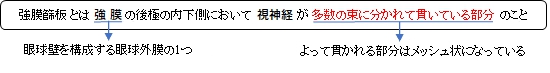
「強膜篩板は網膜から視神経が強膜を貫く部分。 ①強膜の後極近く(後極の内側下側)で視神経が強膜を貫く部分。強膜は篩状になりその孔を多数の束に分かれた視神経が通る。」
以下は「Wikipedia」の解説文となる。
The nerve fibers forming the optic nerve exit the eye posteriorly through a hole in the sclera that is occupied by a mesh-like structure called the lamina cribrosa. It is formed by a multilayered network of collagen fibers that extend from the scleral canal wall. The nerve fibers that comprise the optic nerve run through pores formed by these collagen beams. In humans, a central retinal artery is located slightly off-center in the nasal direction.
【 語 句 】
・collagen fibers: ・comprise:含む、構成する ・pore:細穴 ・central retinal artery:網膜中心動脈
The lamina cribrosa is thought to help support the retinal ganglion cell axons as they traverse the scleral canal. Being structurally weaker than the much thicker and denser sclera, the lamina cribrosa is more sensitive to changes in the intraocular pressure and tends to react to increased pressure through posterior displacement. This is thought to be one of the causes of nerve damage in glaucoma, as the displacement of the lamina cribrosa causes the pores to deform and pinch the traversing nerve fibers and blood vessels.[2]
【 語 句 】
・retinal ganglion cell:網膜神経節細胞 ・axon:軸索 ・scleral canal:角膜孔 ・intraocular pressure:眼圧 ・displacement:転移、置換 ・glaucoma:緑内障 ・deform:変形する ・pinch:締め付ける
■ 写真やイラストを掲載しているサイト ■
・ イラストや写真を掲載しているサイト-Ⅰ
・ イラストや写真を掲載しているサイト-Ⅱ
・ イラストや写真を掲載しているサイト-Ⅲ
・ イラストや写真を掲載しているサイト-Ⅳ
・ イラストや写真を掲載しているサイト-Ⅴ


