【概 要】

・厚みはおよそ50~150㎚(Google Gemin)

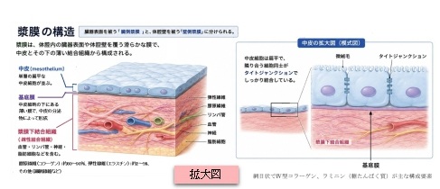
以下は漿膜における基底膜を表した図でとなる。
【構 造】
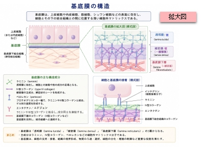
胃底膜は以下の3層構造となる。

【存在する場所】
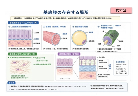
基底膜は主に以下のような場所に存在する。
| |
分類 |
具体的な存在場所 |
その場所での主な役割 |
1 |
体表・管腔の表面
(上皮細胞の下) |
皮膚(表皮と真皮の境界) |
表皮を真皮に強力に繋ぎ止め、外力から守る。 |
消化管(食道、胃、腸など) |
栄養の吸収をコントロールし、病原体の侵入を防ぐ。 |
呼吸器(気管、気管支など) |
吸い込んだ異物を排出する繊毛細胞などの土台となる。 |
2 |
内臓の包膜・空間
(中皮細胞の下) |
漿膜(胸膜、腹膜、心膜) |
臓器の摩擦を減らす中皮の土台。水分の移動を制御。 |
3 |
血管・リンパ管
(内皮細胞の下) |
毛細血管・細動脈など |
血管の構造を保ち、血液成分が外に漏れるのを防ぐ。 |
4 |
特定の臓器・高機能膜
(特殊なフィルター) |
腎臓の糸球体 |
血液をろ過して尿を作る、体内で最も分厚い基底膜。 |
肺胞 |
ガス交換をスムーズにするため、極限まで薄い。 |
5 |
細胞の周囲を包む膜
(外層・外板) |
骨格筋・心筋(筋肉細胞) |
筋肉の激しい収縮による細胞膜の破壊を防ぐ(クッション)。 |
末梢神経(シュワン細胞) |
神経線維を包み込み、神経の保護や再生を助ける。 |
脂肪細胞 |
1つ1つの脂肪細胞の周囲をすっぽり包んで形を保つ。 |
【参考になるサイト】
以下は「Wikipedia」の解説文となる。
The basement membrane, also known as base membrane is a thin, pliable sheet-like type of extracellular matrix that provides cell and tissue support and acts as a platform for complex signalling.[1][2] The basement membrane sits between epithelial tissues including mesothelium and endothelium, and the underlying connective tissue.[3][4]
【Structure】
Normal
histology of the
breast, with basement membrane annotated near center-right.
As seen with the
electron microscope, the basement membrane is composed of two layers, the
basal lamina and the
reticular lamina.
[4] The underlying connective tissue attaches to the basal lamina with
collagen VII anchoring fibrils and
fibrillin microfibrils.
[5]
The basal lamina layer can further be subdivided into two layers based on their visual appearance in electron microscopy. The lighter-colored layer closer to the epithelium is called the
lamina lucida, while the denser-colored layer closer to the connective tissue is called the
lamina densa. The
electron-dense lamina densa layer is about 30–70
nanometers thick and consists of an underlying network of reticular
collagen IV fibrils which average 30 nanometers in diameter and 0.1–2
micrometers in thickness and are coated with the heparan sulfate-rich proteoglycan
perlecan.
[6] In addition to collagen, this supportive matrix contains intrinsic macromolecular components. The lamina lucida layer is made up of
laminin,
integrins,
entactins, and
dystroglycans. Integrins are a key component of
hemidesmosomes which serve to anchor the epithelium to the underlying basement membrane.
To represent the above in a visually organised manner, the basement membrane is organized as follows:
The primary function of the basement membrane is to anchor down the epithelium to its loose connective tissue (the dermis or lamina propria) underneath. This is achieved by cell-matrix adhesions through substrate adhesion molecules (SAMs).
The basement membrane acts as a mechanical barrier, preventing malignant cells from invading the deeper tissues.[7] Early stages of malignancy that are thus limited to the epithelial layer by the basement membrane are called carcinoma in situ.
The basement membrane is also essential for angiogenesis (development of new blood vessels). Basement membrane proteins have been found to accelerate differentiation of endothelial cells.[8]
The most notable examples of basement membranes is the glomerular basement membrane of the kidney, by the fusion of the basal lamina from the endothelium of glomerular capillaries and the podocyte basal lamina,[9] and between lung alveoli and pulmonary capillaries, by the fusion of the basal lamina of the lung alveoli and of the basal lamina of the lung capillaries, which is where oxygen and CO2 diffusion occurs (gas exchange).
As of 2017, other roles for basement membrane include blood filtration and muscle homeostasis.
[1] Fractones may be a type of basement membrane, serving as a
niche for
stem cells
【 語 句 】
・: ・: ・: ・: ・: ・: ・: ・: ・: ・: ・: ・: ・: ・: ・: ・: ・: ・: ・: ・: ・: ・: ・: ・: ・: ・: ・: ・: ・: ・: ・: ・:
■ 写真やイラストを掲載しているサイト ■
・ イラストや写真を掲載しているサイト-Ⅰ
・ イラストや写真を掲載しているサイト-Ⅱ
・ イラストや写真を掲載しているサイト-Ⅲ