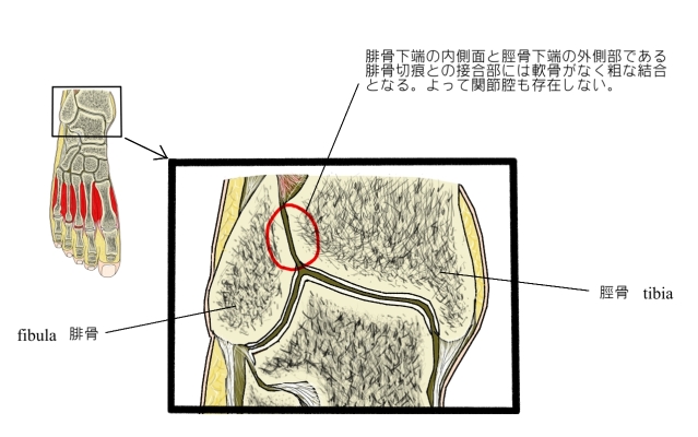
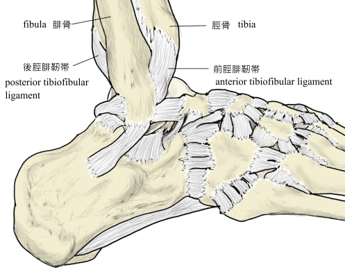
脛腓靭帯結合 (けいひじんたいけつごう、英:talofibular syndesmosis) 脛骨下端の外側部である腓骨切痕と腓骨下端の内側面との結合は、特別な関節腔を持たず粗な面で接している。そしてこの結合面を、前部は前脛腓靭帯で、そして後部は後脛腓靭帯で補強している。これらの結合方法を脛腓靭帯結合と呼ぶ。 「船戸和弥のホームページ」では以下のような解説が見られる。 「この部分に関節腔はないが、距腿関節の関節腔が上方にのびてくることがある。」 右足首(断面) 右足首周辺(外側面) また、「Wikipedia」では、「下腿骨間膜」も補強する靭帯の一つにしている。以下が解説文となる。 「The distal tibiofibular joint (tibiofibular syndesmosis) is formed by the rough, convex surface of the medial side of the distal end of the fibula, and a rough concave surface on the lateral side of the tibia. Below, to the extent of about 4 mm. these surfaces are smooth, and covered with cartilage, which is continuous with that of the ankle-joint. The ligaments are:・Anterior ligament of the lateral malleolus ・Posterior ligament of the lateral malleolus ・Interosseous membrane of leg」 【イラスト・写真を掲載しているサイト】 ・イラストを掲載しているサイトⅠ(靭帯のCG) ・イラストを掲載しているサイトⅡ(靭帯のイラスト) ・イラストを掲載しているサイトⅢ(冠状断面)