「 船戸和弥のHP 」では以下のように解説している。
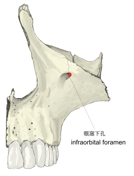
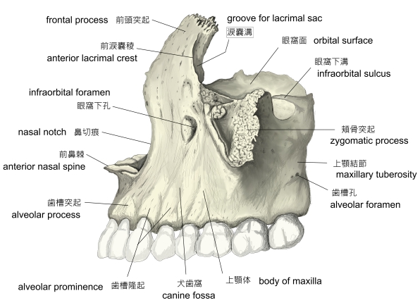
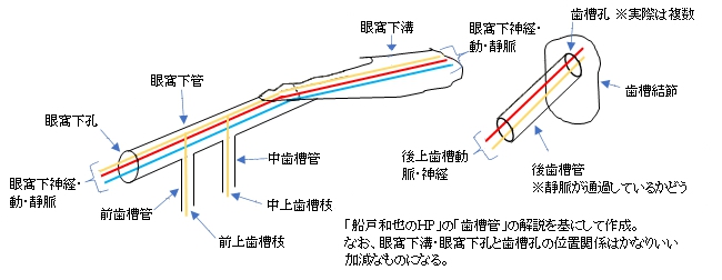
「上顎体下縁は明瞭な境なしに歯槽突起に移る。眼窩下縁の下約0.5-1.0cmに眼窩下孔がある。これは眼窩面の眼窩下溝につづく眼窩下管の顔面に開く口である。」
以下は「Wikipedia」の解説文となる。
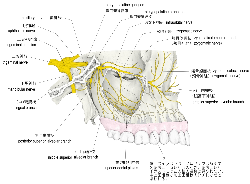
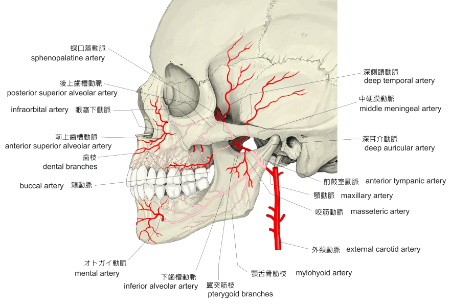
In human anatomy, the infraorbital foramen is an opening in the maxillary bone of the skull located below the infraorbital margin of the orbit. It transmits the infraorbital artery and vein, and the infraorbital nerve, a branch of the maxillary nerve. It is typically 6.10 to 10.9 mm (0.240 to 0.429 in) from the infraorbital margin.
【Structure】
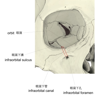
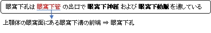
Forming the exterior end of the infraorbital canal, the infraorbital foramen communicates with the infraorbital groove, the canal's opening on the interior side.
The ramifications of the three principal branches of the trigeminal nerve—at the supraorbital, infraorbital, and mental foramen—are distributed on a vertical line (in anterior view) passing through the middle of the pupil. The infraorbital foramen is used as a pressure point to test the sensitivity of the infraorbital nerve. Palpation of the infraorbital foramen during an extraoral examination or an administration of a local anesthetic agent will cause soreness to the area.
【 語 句 】
・maxillary bone:上顎骨 ・infraorbital margin:眼窩縁 ・orbit:眼窩 ・infraorbital artery:眼窩下動脈 ・infraorbital nerve:眼窩下神経 ・maxillary nerve:上顎神経 ・infraorbital groove:眼窩下溝 ・ramification: ・trigeminal nerve: ・mental foramen: ・distribute: ・pupil: ・palpation: ・administration:投与 ・local anesthetic agent:局所麻酔薬 ・soreness:ひりひりすること
■ 写真やイラストを掲載しているサイト ■
・ イラストや写真を掲載しているサイト-Ⅰ
・ イラストや写真を掲載しているサイト-Ⅱ
・ イラストや写真を掲載しているサイト-Ⅲ
・ イラストや写真を掲載しているサイト-Ⅳ