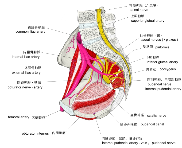
陰部神経管 ( いんぶしんけいかん、 英: pudendal canal ) 陰部神経管とは ・上で「骨盤内の内閉鎖筋と坐骨の間?」と記しているが、インターネットで画像検索をしてみると以下の2つのパターンが見られる。 1 . 骨盤を内側から見た場合、陰部神経管が内閉鎖筋の下部(内閉鎖筋と坐骨の間)に位置するように描かれ ているもの。 ( 下の左のイラスト ) 2 . 骨盤を内側から見た場合、陰部神経管が内閉鎖筋の縁(坐骨の付着部付近)付近の筋膜に包まれるように 描かれているもの。 ( 下の右のイラスト ) 以下は「船戸和弥のホームページ」の解説文となる。 「陰部神経管はアルコック管とも呼ばれる。内閉鎖筋の内側に沿って位置するトンネル状構造で、内閉鎖筋膜にかこまれた形で存在する。内陰部動・静脈および陰部神経が通る。」 また、「 Wikipedia 」では以下のように解説している。 「 The pudendal canal ( also called Alcock's canal ) is an anatomical structure in the pelvis through which the internal pudendal artery, internal pudendal veins, and the pudendal nerve pass. 【 structure 】 The pudendal canal is formed by the fascia of the obturator internus muscle, or obturator fascia. It encloses the following: ・ Internal pudendal artery ・ Internal pudendal veins ・ Pudendal nerve These vessels and nerve cross the pelvic surface of the obturator internus. 」 【 参考になるサイト 】 ・ イラストや写真を掲載しているサイト-Ⅰ ・ イラストや写真を掲載しているサイト-Ⅱ ・ イラストや写真を掲載しているサイト-Ⅲ ・ イラストや写真を掲載しているサイト-Ⅳ