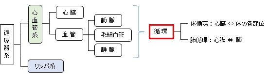
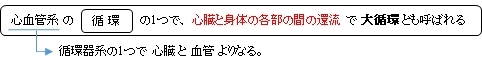
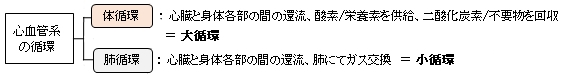
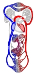
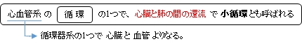
循環器系における心血管系の「循環」とは血液の循環を意味する。血液は全身の細胞に酸素や栄養素を送り、細胞が活動する過程で発生した不要物を回収するという生命を維持するうえで欠かせない働きを担っている。その循環は、大きく心臓と身体の各部の間を還流する体循環(/大循環)と心臓と肺の間を還流する肺循環(/小循環)とに分けて考えることができる。
以下は「 日本人体解剖学 」の解説文となる。
「 心血管系には、心臓と身体各部の間を還流する体循環(大循環)と心臓と肺の間を還流する肺循環(小循環)の2系統があるが、心臓を間に1つの閉鎖性回路を形成している。 」

体循環とは

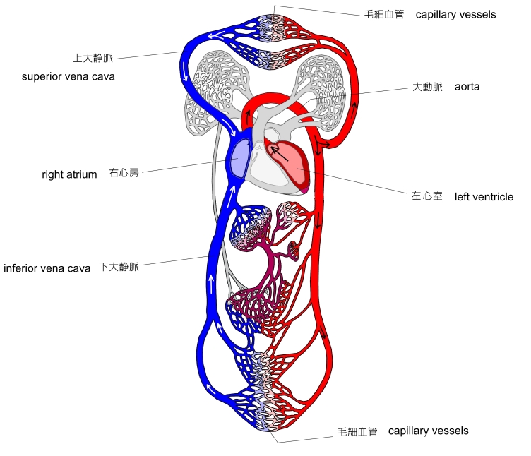
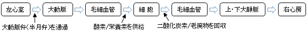
体循環は以下のような経過をたどる。
体循環
模型図
|
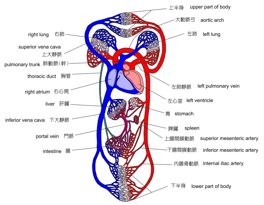
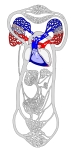
全身の循環器系
模型図
|
|
|
|
以下は「 Wikipedia 」の解説文となる。
「 Systemic circulation is the portion of the cardiovascular system which transports oxygenated blood away from the heart through the aorta from the left ventricle where the blood has been previously deposited from pulmonary circulation, to the rest of the body, and returns oxygen-depleted blood back to the heart. 」
【 語 句 】
・ portion : 部分 ・ oxygenated : 酸素を含んだ ・ aorta : 大動脈 ・ left ventricle : 左心室
・ pulmonary circulation : 肺循環 ・deplete : 使いつくす
また、ウィキペディアの日本語版では以下のように解説している。
「 体循環( たいじゅんかん、Systemic circulation、別名:大循環 )とは、左心室→大動脈→組織→大静脈→右心房の経路に循環すること。体循環と肺循環の血液量は、つまり、左心室と右心室の拍出量は、同じであるようにおもえるが、体循環と肺循環の間には一部分に短縮(生理的シャント)が存在する。 例えば、気管支動脈肺動脈に、冠状動脈の一部分が左心房に還流する。そうすると、正常でも約2%の体循環血は、右心系に戻れない。この、短縮された量は、病的な場合には異常的に増す。 体循環の収縮期血圧は、肺循環の5倍の圧が加わっている。これは、体血管抵抗が、肺血管抵抗の5倍だからである。 」
【 イラスト掲載サイト 】
・ イラストや写真を掲載しているサイト-Ⅰ
・ イラストや写真を掲載しているサイト-Ⅱ
・ イラストや写真を掲載しているサイト-Ⅲ
・ イラストや写真を掲載しているサイト-Ⅳ

肺循環とは
「 人体のしくみと病気 」では吸気と呼気の各ガスの割合を以下のようにしている。
・ 吸 気 : 窒素 ( 79 % )、 酸素 ( 21 % )、 二酸化炭素 ( 0.03 % )
・ 呼 気 : 窒素 ( 79 % )、 酸素 ( 16 % )、 二酸化炭素 ( 4 % )
また、ウィキペディアでは、肺循環の働きとしてガス交換の他に以下を挙げている。
血液ろ過作用 :「 血栓などは肺循環において捕捉されたのち融解されたりして除去される。」
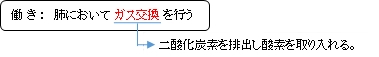
肺循環は以下のような経過をたどる。
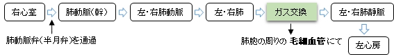
・ 肺において二酸化炭素を排出し酸素を取り入れることを 外呼吸 という。


以下は「 ウィキペディア 」の解説文となる。
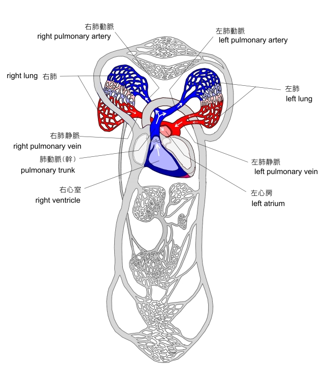
「 肺循環は心臓の肺動脈弁を越えたところから始まりすぐに左右2つの肺動脈に分岐し、気管支と並行して枝分かれしながら二次小葉の中心に達し毛細血管として肺胞壁を取り囲む。ここで酸素を得たのち、合流し小肺静脈として小葉間隔壁を走り左右の肺で上下2本、計4本の肺静脈になり左心房に流れ、肺循環を終了する。肺循環時間は4~6秒でこのうち赤血球が肺毛細血管を通過する時間は安静時で0.75秒で、運動時には0.25秒まで短縮する。 」
肺循環
模型図
|
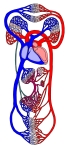
全身の循環器系
模型図
|
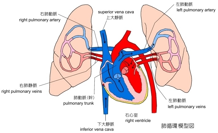
肺循環・模型図-Ⅱ
|
|
|
以下は「 Wikipedia 」の解説文となる。
「 The pulmonary circulation is the portion of the circulatory system which carries deoxygenatedblood away from the right ventricle of the heart, to the lungs, and returns oxygenated blood to the left atrium and ventricle of the heart.[1] The term pulmonary circulation is readily paired and contrasted with the systemic circulation. The vessels of the pulmonary circulation are the pulmonary arteries and the pulmonary veins.
A separate system known as the bronchial circulation supplies oxygenated blood to the tissue of the larger airways of the lung.
The earliest human discussions of pulmonary circulation date back to Egyptian times. Human knowledge of pulmonary circulation grew gradually over centuries, and scientists Ibn al-Nafis, Michael Servetus, and William Harvey provided some of the first accurate descriptions of this process. 」
【 語 句 】
・ portion : 部分 ・ deoxygenated : 脱酸素した( = 酸素が少なくなった ) ・ right ventricle : 右心室
・ left atrium : 左心房 ・ bronchial circulation : 気管支循環
【 イラスト掲載サイト 】
・ イラストや写真を掲載しているサイト-Ⅰ
・ イラストや写真を掲載しているサイト-Ⅱ
・ イラストや写真を掲載しているサイト-Ⅲ
・ イラストや写真を掲載しているサイト-Ⅳ
・ イラストや写真を掲載しているサイト-Ⅴ

■ 門(静)脈 : portal vein ■


目 的 : 胃や腸などの消化管で体内に取り入れられた栄養素を体内で利用しやすい形に変える働き(代謝)をしている。

以下は「Rauber-Kopsch解剖学」の解説文となる。
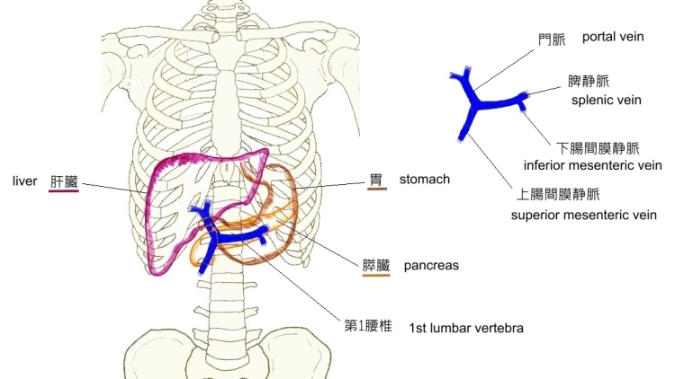
「門脈の幹は6~8cmの長さで,膵頭のうしろで上腸間膜静脈と脾静脈とが合することによって始ま り,斜めに右上方に向かって肝門にいたる」

「 Wikipedia 」には以下のような解説文が見受けられる。
「 Approximately 75% of total liver blood flow is through the portal vein, with the remainder coming from the hepatic artery proper. 」
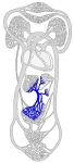
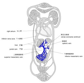
門脈・模型図
|
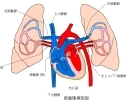
門脈系
|
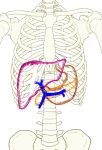
門脈の位置
|
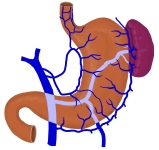
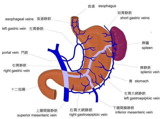
胃周辺の静脈
|
全身の循環器系
模型図
|
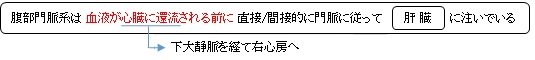
以下は「 日本人体解剖学 」の解説文となる。 ※ 図は私が作成したもの。
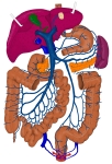
「 毛細血管から静脈になり、再び毛細血管になる場合の静脈を門(静)脈という。腹部内臓、とくに胃・腸・脾臓・膵臓・胆嚢などから集まった静脈血は、直接下大静脈にそそぐことなく、門(静)脈をへて肝臓中に入る。門(静)脈の枝は肝動脈の枝とともに毛細血管(類洞)となった後、再び静脈となり、肝静脈として下大静脈にそそぐ。下錐体にも下垂体門脈系が存在する。」
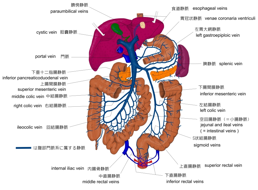
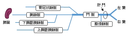
「 門脈は、脾臓・胃・腸管および膵臓・胆嚢からの血液をあつめこれを肝臓に導くもので、脾静脈、上腸間膜静脈、下腸間膜静脈の3主根および胃の小弯を走る胃冠状静脈、胆嚢からくる胆嚢静脈の2副根からできる(腹壁からの臍傍静脈も門脈に入る)
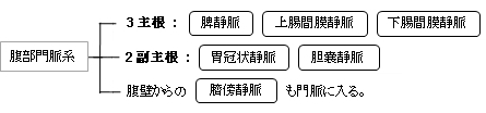
腹部内臓から起こる前記の静脈のうち、胃冠状静脈と下腸間膜静脈はまず脾静脈に集まり、これに上腸間膜静脈が加わって門脈を構成する(一般に、胃冠状静脈は直接門脈に開口する例が多い)。このようにして、門脈の幹は膵頭の後側に始まり、上右方に向かい十二指腸上部後方に進み、次に肝十二支腸間膜(総胆管と肝動脈との間でその後ろに位置する)を通って肝門に達し、ここで右枝および左枝に分かれて肝臓の左・右両葉中に入り分枝する。」
「 プロメテウス解剖学アトラス:頚部/胸部/腹部・骨盤部 」では「門脈の枝」として以下の静脈を挙げている。
・ 膵十二指腸静脈 ・ 膵静脈 ・ 右胃大網静脈 ・ 空腸静脈 ・ 回腸静脈 ・ 回結腸静脈 ・ 右結腸静脈
・ 中結腸静脈
・ 左結腸静脈 ・ S状結腸静脈 ・ 上直腸静脈
・ 左胃大網静脈 ・ 膵静脈 ・ 短胃静脈
・ 胆嚢静脈 ・ 左胃静脈と食道静脈 ・ 右胃静脈 ・ 後上膵十二指腸静脈 ・ 幽門前静脈 ・ 臍傍静脈
以下は「 Wikipedia 」の解説文となる。
「The portal vein or hepatic portal vein is a blood vessel that carries blood from the gastrointestinal tract, gallbladder, pancreas and spleen to the liver. This blood contains nutrients and toxins extracted from digested contents. Approximately 75% of total liver blood flow is through the portal vein, with the remainder coming from the hepatic artery proper. The blood leaves the liver to the heart in the hepatic veins.
The portal vein is not a true vein, because it conducts blood to capillary beds in the liver and not directly to the heart. It is a major component of the hepatic portal system, one of only two portal venous systems in the body – with the hypophyseal portal system being the other.
The portal vein is usually formed by the confluence of the superior mesenteric and splenic veinsand also receives blood from the inferior mesenteric, gastric, and cystic veins.
Conditions involving the portal vein cause considerable illness and death. An important example of such a condition is elevated blood pressure in the portal vein. This condition, called portal hypertension, is a major complication of cirrhosis.
【 語 句 】
・ gastrointestinal tract : 消化管 ・ gallbladder : 胆嚢 ・ toxin : 毒素 ・ extrac : 取り出す
・ digested content : 消化物 ・ capillary bed : 毛細血管床 ・ hypophyseal : 下垂体 ・ confluence : 合流
・ cystic vein : 胆嚢静脈 ・ portal hypertension : 門脈圧亢進症 ・ complication : 合併症 ・ cirrhosis : 肝硬変
【 structure 】
Measuring approximately 8 cm (3 inches) in adults,[1] the portal vein is located in the right upper quadrant of the abdomen, originating behind the neck of the pancreas.[2]
In most individuals, the portal vein is formed by the union of the superior mesenteric vein and the splenic vein.[3] For this reason, the portal vein is occasionally called the splenic-mesenteric confluence.[2] Occasionally, the portal vein also directly communicates with the inferior mesenteric vein, although this is highly variable. Other tributaries of the portal vein include the cystic and gastric veins.[4]
| Tributaries of the hepatic portal vein[4] |
|
Immediately before reaching the liver, the portal vein divides into right and left. It ramifies further, forming smaller venous branches and ultimately portal venules. Each portal venule courses alongside a hepatic arteriole and the two vessels form the vascular components of the portal triad. These vessels ultimately empty into the hepatic sinusoids to supply blood to the liver.[4]
・ Portacaval anastomoses
The portal venous system has several anastomoses with the systemic venous system. In cases of portal hypertension these anastamoses may become engorged, dilated, or varicosed and subsequently rupture.
・ Accessory hepatic portal veins
Accessory hepatic portal veins are those veins that drain directly into the liver without joining the hepatic portal vein. These include the paraumbilical veins as well as veins of the lesser omentum, falciform ligament, and those draining the gallbladder wall.[2]
【 語 句 】
・ tributary : 支流 ・ ramify : 分岐する ・ ultimately : 最後には ・ venule : 細静脈 ・ hepatic : 肝臓の
・ arteriole : 小動脈 ・ vascular : 導管の ・ triad : 三連構造 ・ sinusoid : 類洞 ・Portacaval : 門脈大静脈の
・ anastomose : 吻合 ・ engorge : 充血させる ・ dilate:ふくらませる ・ varicosed:静脈瘤の
・subsequently :続いて
・rupture : 破裂/ヘルニア ・ paraumbilical vein : 臍傍静脈 ・ lesser omentum : 小網
・ falciform ligament : 鎌状靭帯
【 function 】
The portal vein and hepatic arteries form the liver's dual blood supply. Approximately 75% of hepatic blood flow is derived from the portal vein, while the remainder is from the hepatic arteries.[2]
Unlike most veins, the portal vein does not drain into the heart. Rather, it is part of a portal venous system that delivers venous blood into another capillary system, the hepatic sinusoids of the liver. In carrying venous blood from the gastrointestinal tract to the liver, the portal vein accomplishes two tasks: it supplies the liver with metabolic substrates and it ensures that substances ingested are first processed by the liver before reaching the systemic circulation. This accomplishes two things. First, possible toxins that may be ingested can be detoxified by the hepatocytes before they are released into the systemic circulation.
Second, the liver is the first organ to absorb nutrients just taken in by the intestines. After draining into the liver sinusoids, blood from the liver is drained by the hepatic vein. 」
【 語 句 】
・ venous : 静脈の ・ capillary : 毛細血管 ・ metabolic substrate : 代謝基質 ・ ingest : 摂取する ・ detoxify : 解毒する ・ hepatocyte : 幹細胞
【 イラスト掲載サイト 】
・ イラストや写真を掲載しているサイト-Ⅰ
・ イラストや写真を掲載しているサイト-Ⅱ
・ イラストや写真を掲載しているサイト-Ⅲ
・ イラストや写真を掲載しているサイト-Ⅳ

|