尺骨動脈 とは
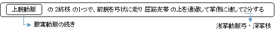
・「 その経過は肘窩と尺骨茎状突起とを結ぶゆるい弓形線を描く。」( 日本人体解剖学 )
・「 この動脈はたいてい橈骨動脈より弱くて,前腕の内側を遠位に向かって走っている.」( 船戸和弥のホームページ )
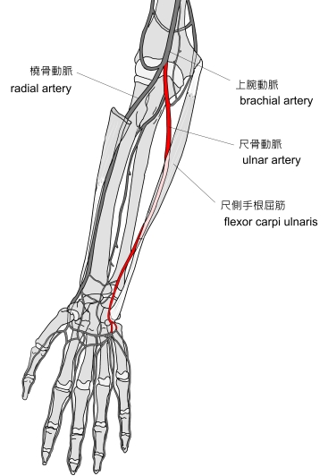
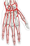
右手( 前 面 ) |

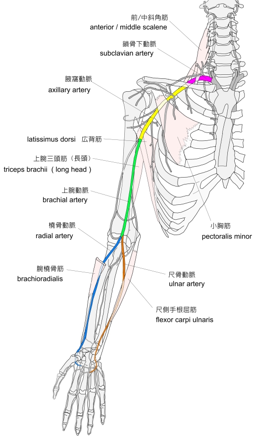
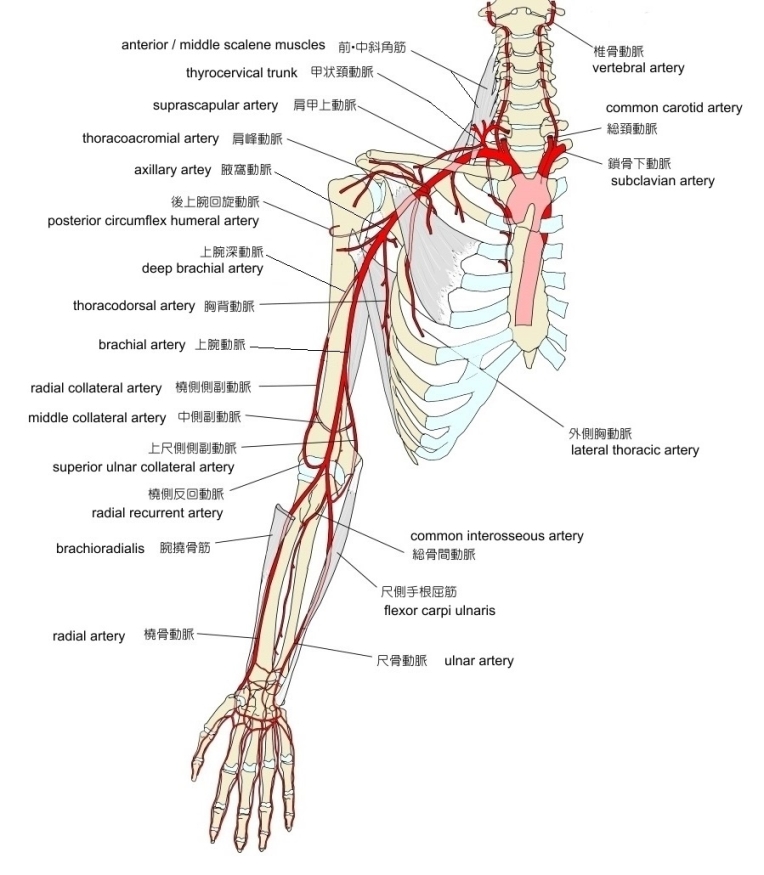
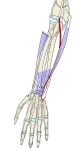
右上肢( 前 面 ) |
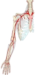
右上肢( 前 面 ) |
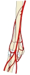
右肘窩周辺 |
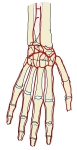
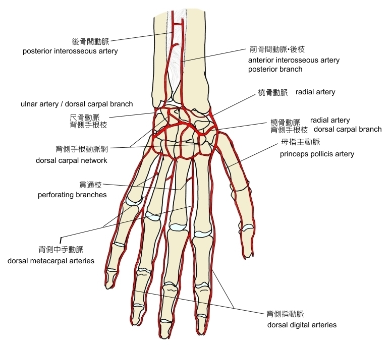
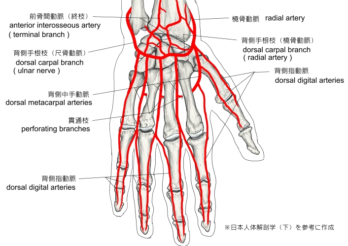
右手( 背 面 ) |
右手( 背 面 )
|
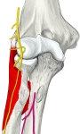
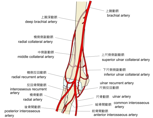
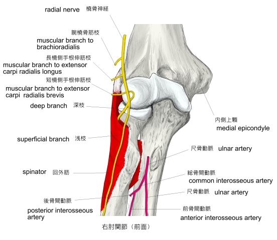
右肘関節(前面)
|
|
|
|
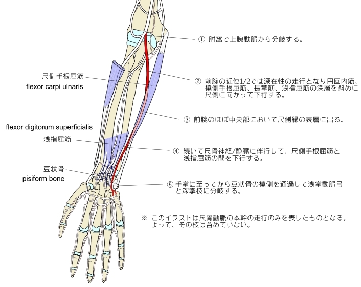
1 . 肘窩で上腕動脈から分岐する。( 肘関節の少し遠位の位置? )
( 分岐部あたりでは橈骨動脈よりも太い )
2 . 前腕の近位1/2では深在性の走行となり、円回内筋・橈側手根屈筋・長掌筋・浅指屈筋の深層を斜め尺側に向かって下行する。
3 . 前腕のほぼ中央部において尺側縁の表層に出る。
4 . 続いて尺骨神経/静脈に伴行して、尺側手根屈筋と浅指屈筋との間を下行する。
5 . 手掌に至ってから豆状骨の橈側を通過して、浅掌動脈弓と深掌枝に分岐する。

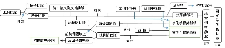
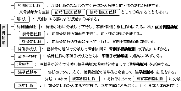
以下は尺側動脈の枝を簡単に表したものとなる。 ※ 参考 :「 日本人体解剖学 」


以下は「 Rauber-Kopsch 解剖学 」の解説文となる。
「 R. Quainが観察したところでは尺骨動脈の起始は13例中およそ1例の割りで変異を示していた.この場合には尺骨動脈が腋窩動脈から出ることよりも上腕動脈から出ていることがずっと多く,しかも変異の見られる回数は正常に出る場所からその起始が遠ざかるほどますます減少していた. 前腕における尺骨動脈の位置は橈骨動脈の場合よりも変化が多い.尺骨動脈が正常の場所で始まっているときにはその位置にあまり変化が多くない.しかしこの動脈が正常には尺側手根屈筋の腱に接しているが,そうなっていないでこの筋から離れて走っていることがしばしばある.--尺骨動脈が高位で始まる場合はほとんど例外なくこれが上腕骨の尺側上顆に始まる諸筋の上を越えている,たいていこの動脈は上腕の筋膜に被われてその下にあるが,これがもっと浅くて皮下を通っていることがあり,その全経過を通じて皮下の表層を走り続けることがあり,あるいは後になって筋膜の下にはいりこみ,さらに遠位でこの動脈の正常な位置になるということもある.
E. Zuckerkandl (1896)によると多くの例で(79%)尺骨動脈に2本の深掌枝があり,そのうち遠位のものが普通にはずっと強大である.近位の枝が欠けていることは決してない(100例をしらべて).しかし遠位の枝は21%に欠けている.近位の枝は豆状骨の近くで始まるが,遠位の枝は浅掌枝が曲がって浅掌動脈弓になるところから出ている(図658).」

以下は「 Wikipedia 」の解説文となる。
「 The ulnar artery is the main blood vessel, with oxygenated blood, of the medial aspect of the forearm. It arises from the brachial artery and terminates in the superficial palmar arch, which joins with the superficial branch of the radial artery. It is palpable on the anterior and medial aspect of the wrist.
Along its course, it is accompanied by a similarly named vein or veins, the ulnar vein or ulnar veins.
The ulnar artery, the larger of the two terminal branches of the brachial, begins a little below the bend of the elbow in the cubital fossa, and, passing obliquely downward, reaches the ulnar side of the forearm at a point about midway between the elbow and the wrist. It then runs along the ulnar border to the wrist, crosses the transverse carpal ligament on the radial side of the pisiform bone, and immediately beyond this bone divides into two branches, which enter into the formation of the superficial and deep volar arches.
【 語 句 】
・ superficial palmar arch : 浅掌動脈弓 ・ palpable : 触知できる ・ cubital fossa : 肘窩 ・ obliquely : 斜めに ・ transverse carpal ligament : 横手根靭帯 ・ pisiform bone : 豆状骨 ・ superficial and deep volar arches : 浅・深掌動脈弓
【 Branches 】
Forearm :
・ Anterior ulnar recurrent artery,
・ Posterior ulnar recurrent artery,
・ Common interosseous
( posterior, and recurrent interosseous arteries )
・ palmar carpal branch
・ dorsal carpal branch
Hand :
・ Deep palmar branch
・ superficial palmar arch.
【 Relations 】
In its upper half, it is deeply seated, being covered by the Pronator teres, Flexor carpi radialis, Palmaris longus, and Flexor digitorum superficialis; it lies upon the Brachialis and Flexor digitorum profundus.
The median nerve is in relation with the medial side of the artery for about 2.5 cm. and then crosses the vessel, being separated from it by the ulnar head of the Pronator teres.
In the lower half of the forearm it lies upon the Flexor digitorum profundus, being covered by the integument and the superficial and deep fascia, and placed between the Flexor carpi ulnaris and Flexor digitorum superficialis.
It is accompanied by two venæ comitantes, and is overlapped in its middle third by the Flexor carpi ulnaris; the ulnar nerve lies on the medial side of the lower two-thirds of the artery, and the palmar cutaneous branch of the nerve descends on the lower part of the vessel to the palm of the hand.
【 Wrist 】
At the wrist the ulnar artery is covered by the integument and the volar carpal ligament, and lies upon the Flexor retinaculum of the hand. On its medial side is the pisiform bone, and, somewhat behind the artery, the ulnar nerve.
【 語 句 】
・ Pronator teres : 円回内筋 ・ Flexor carpi radialis : 橈側手根屈筋 ・ Palmaris longus :長掌筋 ・ Flexor digitorum superficialis : 浅指屈筋 ・ Brachialis : 上腕筋 ・ Flexor digitorum profundus : 深指屈筋 ・ integument : 外皮 ・ fascia : 筋膜 ・ venæ comitantes : 伴走する静脈 ・ palmar cutaneous branch : 掌枝? ・ Flexor retinaculum : 屈筋支帯 ・ volar carpal ligament : 掌側手根靭帯 ・ pisiform bone : 豆状骨
【 Peculiarities 】
The ulnar artery varies in its origin in the proportion of about one in thirteen cases ; it may arise about 5 to 7 cm. below the elbow, but more frequently higher, the brachial being more often the source of origin than the axillary.
Variations in the position of this vessel are more common than in the radial. When its origin is normal, the course of the vessel is rarely changed.
When it arises high up, it is almost invariably superficial to the Flexor muscles in the forearm, lying commonly beneath the fascia, more rarely between the fascia and integument.
In a few cases, its position is subcutaneous in the upper part of the forearm, and subaponeurotic in the lower part. 」
【 語 句 】
・ in the proportion of : ~の割合で? ・ axillary : 腋窩動脈? ・ invariably : 常に ・ subcutaneous : 皮下の ・ subaponeurotic : 腱膜下の
【 イラスト掲載サイト 】
・ イラストや写真を掲載しているサイト-Ⅰ
・ イラストや写真を掲載しているサイト-Ⅱ
・ イラストや写真を掲載しているサイト-Ⅲ
・ イラストや写真を掲載しているサイト-Ⅳ

|