大脳動脈輪とは

・ ウィリスの動脈輪 / ウィリス大脳動脈輪 とも呼ばれる。
( 英国の医学者 Thomas Willis ( 1621-1675 )に因む。)
・「 動脈輪の各部の発達には個人差が著しく、完全な輪が形成されないことがある。」(船戸和弥のホームページ )
以下は「 日本人体解剖学 」の解説文となる。
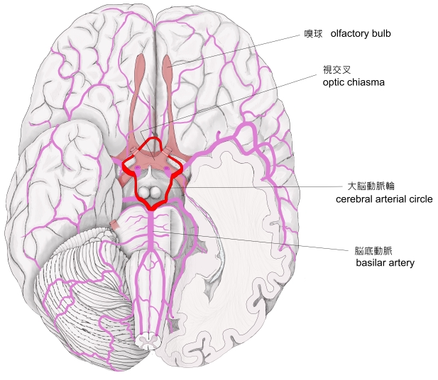
「 大脳動脈輪の位置はトルコ鞍の周囲にあたり、視神経交叉、中央に下垂体漏斗、視床下部の腹側面(灰白隆起および乳頭体)を囲む。 」
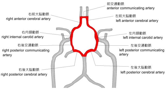
【 構成動脈 】
以下が大脳動脈輪を構成する動脈の一覧となる。
前 方 |
前交通動脈 |
anterior communicating artery |
側 方 |
前 部 |
左・右前大脳動脈 |
left / right anterior cerebral aterty |
後 部 |
左・右内頚動脈 |
left / right internal carotid artery |
左・右後交通動脈 |
left / right posterior communicating artery |
後 方 |
左・右後大脳動脈 |
left / right posterior cerebral artery |

「船戸和弥のホームページ」では以下のように解説している。
「ウィリスの動脈輪ともよばれる。大脳動脈輪は脳底部において、内頸動脈と椎骨動脈の枝が連絡して形成された輪状ないし六角形の動脈吻合である。
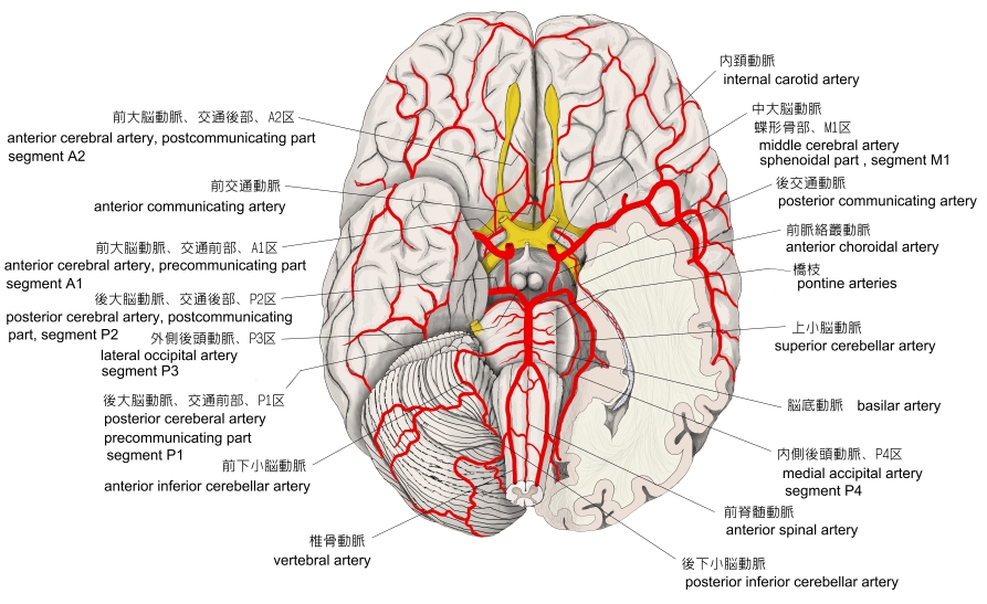
構成にあずかる動脈は、内頚動脈側では前大脳動脈、左右の前大脳動脈を連絡する前交通動脈、中大脳動脈、椎骨動脈側では後大脳動脈、そして中大脳動脈と後大脳動脈を連絡する後交通動脈であり、それらが視神経交叉、下垂体漏斗部、乳頭体、後有孔質などを取り囲む動脈輪を形成する。大脳動脈はすべてこの動脈輪を介して出るということができる。大脳動脈輪は、脳のいろいろな場所へ血液を均等に分配すると言われているが、正常では血圧が等しいので大脳動脈輪の左側と右側との間で血液の交換はほとんど行われない。大脳動脈輪と主要な大脳動脈から2種類の枝、すなわち中心枝と皮質枝が出る。中心枝は、大脳動脈輪と主要な大脳動脈の近位部からでて脳の実質内に入り込み、脳の深部の組織に血液を供給する。前脈絡総動脈と後脈絡叢動脈は、それぞれ内頚動脈の枝と後大脳動脈の枝として出るが、ともにこの中心枝のグループにいれられている。脳内に侵入した血管、とくに中心枝は、他の動脈と吻合しないといわれていて、終動脈とよばれる。人脳では終動脈は存在しないが、大きい血管に突然閉塞が起こると、これらの小動脈の吻合だけでは必要な血液供給を十分に維持することができない。一方皮質枝は、それぞれの主要な大脳動脈から分岐して、軟膜内を通り大脳皮質の広い領域に多数の枝をだしながら、脳表面で自由に吻合して動脈叢を形成する。この動脈叢より分視した小さな動脈は、大脳表面から皮質内にほとんど直角に入り込み、いろいろな深さに達する。動脈輪の各部の発達には個人差が著しく、完全な輪が形成されないことがある。英国の医学者Thomas Willis (1621-1675)により、1664年に発表された。 」
以下は「 Wikipedia 」の解説文となる。
「 The circle of Willis ( also called Willis' circle, loop of Willis, cerebral arterial circle, and Willis polygon ) is a circulatory anastomosis that supplies blood to the brain and surrounding structures. It is named after Thomas Willis (1621–1675), an English physician.[1]
【 Structure 】
The circle of Willis is a part of the cerebral circulation and is composed of the following arteries :[2]
The middle cerebral arteries, supplying the brain, are not considered part of the circle.
【 origin of arteries 】
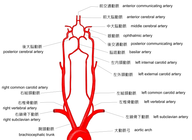
The left and right internal carotid arteries arise from the left and right common carotid arteries.
The posterior communicating artery is given off as a branch of the internal carotid artery just before it divides into its terminal branches - the anterior and middle cerebral arteries. The anterior cerebral artery forms the anterolateral portion of the circle of Willis, while the middle cerebral artery does not contribute to the circle.
The right and left posterior cerebral arteries arise from the basilar artery, which is formed by the left and right vertebral arteries. The vertebral arteries arise from the subclavian arteries.
The anterior communicating artery connects the two anterior cerebral arteries and could be said to arise from either the left or right side.
All arteries involved give off cortical and central branches. The central branches supply the interior of the circle of Willis, more specifically, the Interpeduncular fossa. The cortical branches are named for the area they supply. Since they do not directly affect the circle of Willis, they are not dealt with here.
【 variation 】
Considerable anatomic variation exists in the circle of Willis. Based on a study of 1413 brains, the classic anatomy of the circle is only seen in 34.5% of cases.[3] In one common variation the proximal part of the posterior cerebral artery is narrow and its ipsilateral posterior communicating artery is large, so the internal carotid artery supplies the posterior cerebrum ; this is known as a fetal posterior communicating cerebral artery. In another variation the anterior communicating artery is a large vessel, such that a single internal carotid supplies both anterior cerebral arteries; this is known as an azygos anterior cerebral artery.
【 function 】
The arrangement of the brain's arteries into the circle of Willis creates redundancy (analogous to engineered redundancy) for collateral circulation in the cerebral circulation. If one part of the circle becomes blocked or narrowed (stenosed) or one of the arteries supplying the circle is blocked or narrowed, blood flow from the other blood vessels can often preserve the cerebral perfusion well enough to avoid the symptoms of ischemia.[4]」
【 語 句 】
・ basilar artery : 脳底動脈 ・ subclavian arteries : 鎖骨下動脈 ・ cortical : 皮層の ・ specifically : 具体的に ・ Interpeduncular fossa : 脚間窩 ・ dealt with : 取り扱う ・ proximal : 近い方の ・ipsilateral : 同側の ・ azygos anterior cerebral artery : 奇前大脳動脈 ・ redundancy : 余分 ・ analogous : 類似して ・ perfusion : 潅流 ・ ischemia : 血流不全
【 イラスト掲載サイト 】
・ イラストや写真を掲載しているサイト-Ⅰ
・ イラストや写真を掲載しているサイト-Ⅱ
・ イラストや写真を掲載しているサイト-Ⅲ
・ イラストや写真を掲載しているサイト-Ⅳ
・ イラストや写真を掲載しているサイト-Ⅴ

|