脳底動脈とは


「 日本人体解剖学 」には以下のような解説文が見られる。
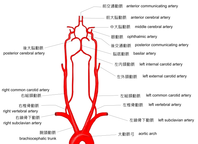
「 左右の椎骨動脈は斜台と延髄との間を上内側方に走り、延髄と橋の境で左右の椎骨動脈が合し脳底動脈を作る。」

以下は脳底動脈の枝を簡単に表した図となる。


以下は「 Wikipedia 」の解説文となる。
「 In human anatomy, the basilar artery is one of the arteries that supplies the brain with oxygen-rich blood.
The two vertebral arteries and the basilar artery are sometimes together called the vertebrobasilar system, which supplies blood to the posterior part of the circle of Willis and joins with blood supplied to the anterior part of the circle of Willis from the internal carotid arteries.
【 Structure 】
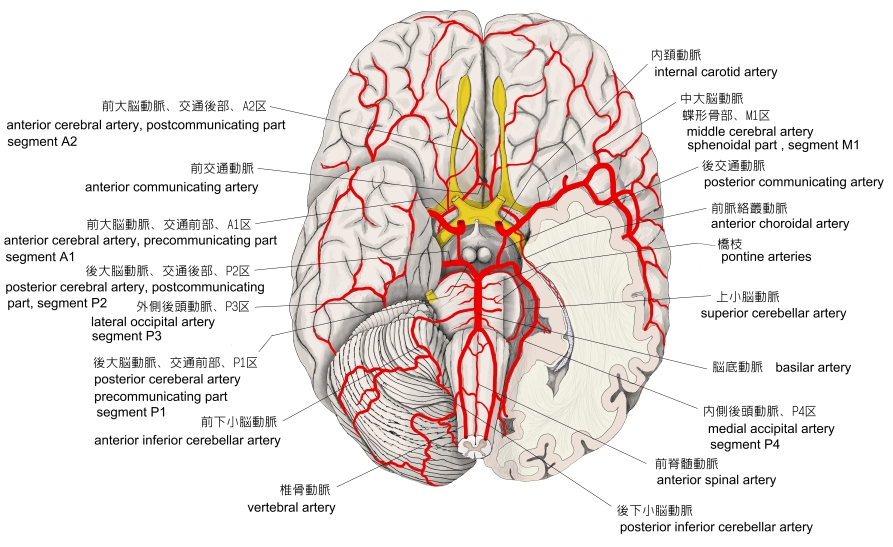
The basilar artery arises from the confluence of the two vertebral arteries at the junction between the medulla oblongata and the pons between the VIth cranial nerves.[1]
It ascends superiorly in the basilar sulcus ventral to the pons and divides at the ponto-mesencephalic junction into the paired posterior cerebral arteries close to the pituitary stalk.
Its branches can be divided into two groups :[1]
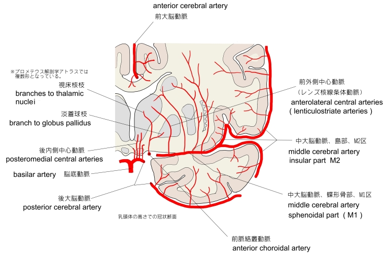
- ■ Paramedian perforating arteries arising either directly from the dorsal surface or from short circumferential arteries running around and into the pons supplying the corticospinal tracts and vital deep nuclei.
- ■ Two or three paired long circumferential branches:
【 語 句 】
・vertebrobasilar system : 椎骨脳底動脈系 ・ circle of Willis : ウィリス大脳動脈輪 ・confluence : 合流 ・ medulla oblongata : 延髄 ・ pons : 橋 ・ basilar sulcus : 脳底溝 ・ pituitary stalk : 下垂体茎 ・ Paramedian : 傍正中の ・ circumferential : 周辺の ・ corticospinal tracts : 皮質脊髄路 ・ vital:nucleus(生命維持に必要な)の複数形 ・ nuclei : 核 ・ labyrinthine artery : 迷路動脈 ・ anterior inferior cerebellar artery: 前下小脳動脈 ・ adjacent : 近隣の ・ hemisphere : 半球 ・ superior cerebellar artery : 上小脳動脈
【 イラスト掲載サイト 】
・ イラストや写真を掲載しているサイト-Ⅰ
・ イラストや写真を掲載しているサイト-Ⅱ
・ イラストや写真を掲載しているサイト-Ⅲ
・ イラストや写真を掲載しているサイト-Ⅳ
・ イラストや写真を掲載しているサイト-Ⅴ

|