前下小脳動脈とは

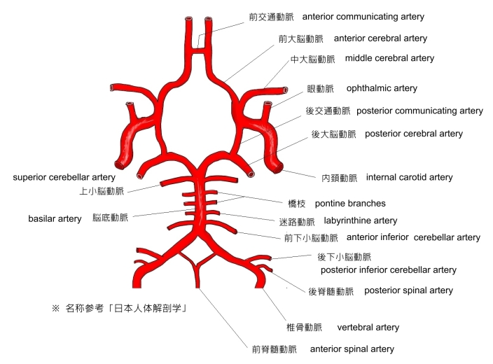
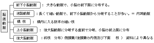
以下は「 日本人体解剖学 」にして脳底動脈の枝を簡単に表した図となる。
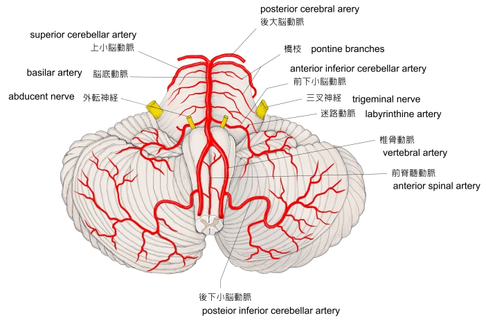
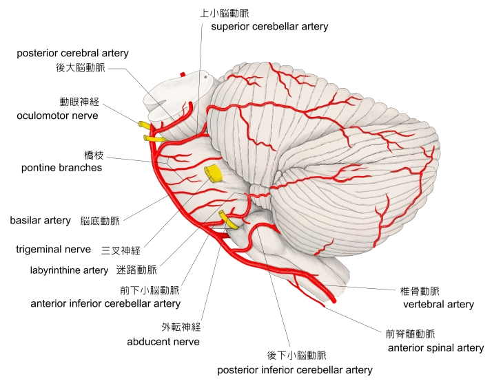
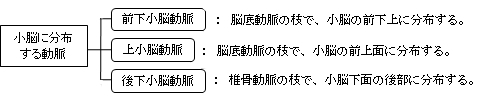
また、以下は小脳に分布する動脈を簡単に表したものとなる。

「 日本人体解剖学 」では「 中央から起こり 」との解説になっているが、「 船戸和弥のホームページ 」では「 尾側部より分岐する 」と解説している。
【 分 布 】
「 日本人体解剖学 」では「小脳の前下面に分布する」と簡単に解説しているだけだが、「 船戸和弥のホームページ 」ではより詳細な解説が見られる。
「 橋被蓋の尾側部に分布した後、尾方に走ってから外側に向かい小脳下面に至る。この動脈は虫部錐体、虫部隆起、片葉、小脳半球の下面の一部に分布する。また深部に穿通する枝は、歯状核の部分とその周囲の白質に分布する。その前下小脳動脈の枝には、第四脳室脈絡叢に分布するものもある。」
・ 橋被蓋の尾側部 ・ 虫部錐体 ・ 虫部隆起 ・ 片葉 ・ 小脳半球の下面の一部
・ 歯状核の部分とその周囲の白質 ・ 第四脳室脈絡叢

以下は「 Wikipedia 」の解説文となる。
「 The anterior inferior cerebellar artery (AICA) is one of three pairs of arteries that supplies blood to the cerebellum.
It arises from the basilar artery on each side at the level of the junction between the medulla oblongata and the pons in the brainstem. It has a variable course, passing backward to be distributed to the anterior part of the undersurface of the cerebellum, anastomosing with both the posterior inferior cerebellar (PICA:) branch of the vertebral artery and the superior cerebellar artery.
It also gives off the internal auditory or labyrinthine artery in most cases; however, the labyrinthine artery can less commonly emerge as a branch of the basilar artery.
The amount of tissue supplied by the AICA is variable, depending upon whether the PICA is more or less dominant, but usually includes the anteroinferior surface of the cerebellum、 the flocculus, middle cerebellar peduncle and inferolateral portion of the pons.[1]」
【 語 句 】
・ basilar artery : 脳底動脈 ・ medulla oblongata : 延髄 ・pons : 橋 ・ brainstem : 脳幹 ・ be distributed to ~ : ~ に分布する ・ posterior inferior cerebellar : 後下小脳動脈 ・ superior cerebellar artery : 上小脳動脈 ・ internal auditory artery : 内耳動脈 ・ labyrinthine artery : 迷路動脈 ・ dominant : 優勢な、高い ・flocculus : 片葉 ・ middle cerebellar peduncle : 中小脳脚
【 イラスト掲載サイト 】
・ イラストや写真を掲載しているサイト-Ⅰ
・ イラストや写真を掲載しているサイト-Ⅱ
・ イラストや写真を掲載しているサイト-Ⅲ
・ イラストや写真を掲載しているサイト-Ⅳ
・ イラストや写真を掲載しているサイト-Ⅴ

|